
Practical, evidence-based recommendations for achieving predictable clinical outcomes in pediatric patients with molar-incisor hypomineralization (MIH), from anesthesia and bonding through restorative care and extraction planning.
1Overcome Anesthetic ResistanceManaging chronic pulpal inflammation and hypersensitivity
Preemptive Analgesia
Administer ibuprofen (10 mg/kg) approximately one hour before the appointment to suppress pulpal prostaglandins and improve anesthetic efficacy.
Optimal Anesthetic Agent
Use 4% articaine instead of lidocaine. Its higher lipid solubility and lower pKa allow better diffusion through inflamed tissue and bone.
Intraosseous (IO) Injections
For mandibular molars resistant to block anesthesia, utilize IO injections (e.g., X-tip system), which bypass cortical bone and achieve profound anesthesia in up to 88.9% of MIH cases.
Photobiomodulation (PBM)
Apply low-level laser therapy (635 nm diode, 60 seconds) at the injection site prior to needle penetration to significantly reduce injection pain and accelerate anesthesia onset.
2Optimize Adhesive BondingDeproteinization to prevent premature restoration failure
Chemical Pre-treatment (Deproteinization)
MIH enamel contains elevated serum albumin that inhibits acid etching. After cavity preparation and before etching, apply 5% sodium hypochlorite (NaOCl) or papain-based gel (Papacarie) for 60 seconds. Rinse thoroughly before proceeding.
Tissue-Driven Margins
Use caries detector dye to confirm all hypomineralized tissue is removed from the cavity periphery. Margins must terminate in sound, dye-free enamel to prevent post-eruptive breakdown (PEB) at the restoration interface.
3Select Appropriate Restorative MaterialsDirect, indirect, and full-coverage options
Direct Restorations
Composite resin is clinically superior to GIC for definitive restorations, with better marginal adaptation and surface texture. Use high-viscosity GIC only as an interim stabilizing material (SMART technique).
Indirect Restorations
For severe structural loss, use resin-infiltrated ceramic CAD/CAM onlays (e.g., Lava Ultimate). Their elastic modulus mimics natural dentin, providing a "cushion effect" against masticatory forces.
Full Coverage
Stainless Steel Crowns (SSCs) remain the gold standard for stabilizing severe, multi-surface defects and eliminating hypersensitivity entirely in young patients.
4Interim Desensitization & StabilizationFor uncooperative patients or highly sensitive erupting molars
Silver Diamine Fluoride (SDF)
Apply 38% SDF to rapidly arrest active decay and desensitize "hot" teeth by forming tubule-blocking silver-protein complexes.
Remineralizing Agents (CPP-ACP)
Prescribe daily at-home use of Casein Phosphopeptide-Amorphous Calcium Phosphate (CPP-ACP) combined with fluoride to enhance mineral density of porous enamel and reduce sensitivity over time.
5Strategic ExtractionsFor poor-prognosis molars with a high restorative burden
Extraction Timing
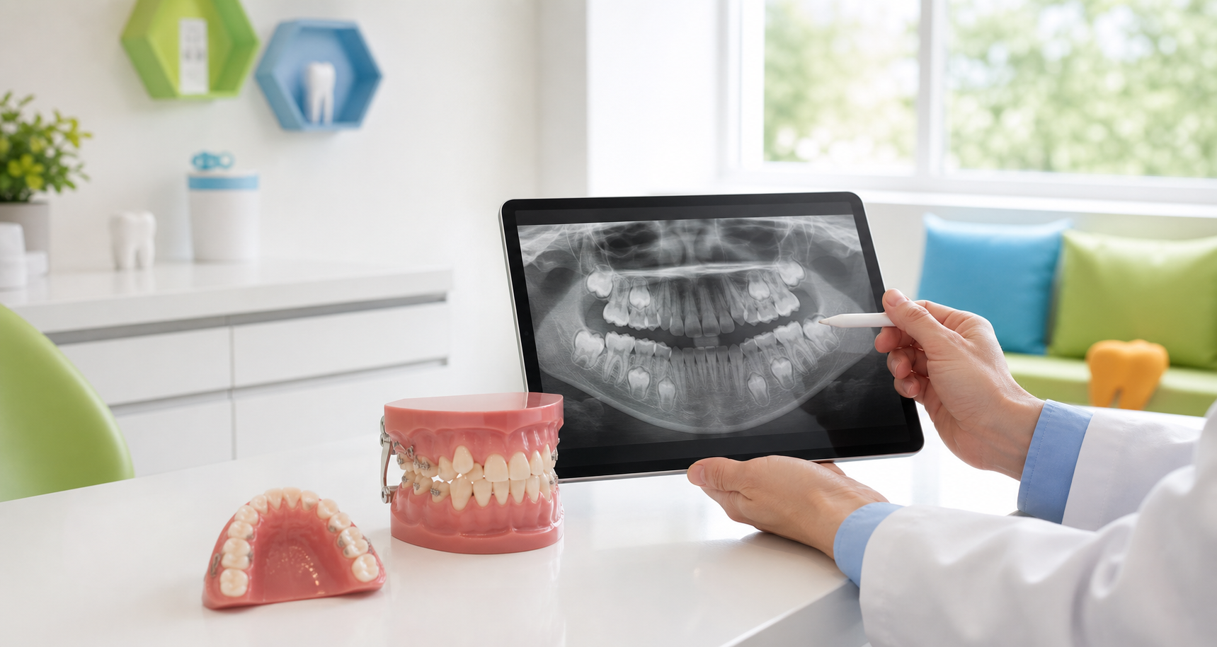
Extract the affected first permanent molar when the developing second permanent molar is at Demirjian Stage E (early root bifurcation), typically between ages 8-10.
Space Closure Predictability
Space closure is highly predictable in the maxilla (85.3%) but less so in the mandible (48.1%). In the mandible, the presence of a developing third molar significantly increases the odds of successful spontaneous closure.
Strategic extraction is a biologically sound alternative when the long-term restorative burden on a first permanent molar is too high. Always assess in the context of the full occlusal development.