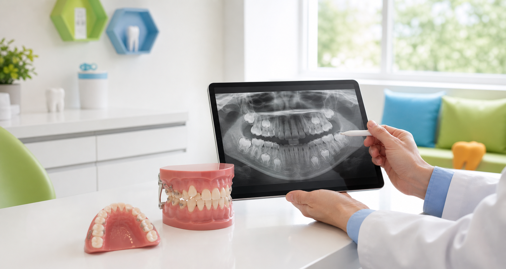
Palatally displaced canines (PDC) are one of the most common dental eruption problems in children. When caught early, a simple interceptive approach — guided by panoramic X-ray measurements — can often redirect the canine to erupt on its own, avoiding surgery entirely.
1The Core Strategy: Extraction of Primary CaninesThe most widely documented interceptive procedure
Extraction Effectiveness
Randomized controlled trials confirm that extraction of the primary canine significantly enhances the probability of spontaneous eruption compared to observation alone — roughly doubling the success rate in most studies.
Predicting Success by Sector (Ericson & Kurol)
If the permanent canine crown overlaps the distal half of the lateral incisor root (Sector 2), extraction yields a 91% success rate. If it has already crossed the midline of the lateral incisor root (Sector 3), success drops to approximately 64%.
2Radiographic Predictors for InterventionUsing panoramic X-rays to decide: intervene, observe, or refer for surgery
The "Golden Window" for Extraction
Interceptive extraction is most beneficial when the alpha angle (α) is between 20° and 30° and the canine is in Sectors 2 or 3. This is the optimal window for spontaneous redirection after extraction.
"Watchful Waiting" Criteria
Observation without intervention is likely to succeed if the alpha angle is less than 20° and the tooth is in Sector 2. These cases often self-correct with no treatment needed.
Unfavorable Indicators — Proceed to Surgery
If the alpha angle exceeds 30° and the tooth is in Sector 4 (overlapping the central incisor), interceptive extraction is often ineffective. Clinicians should proceed directly to surgical exposure and traction.
3Timing & Patient AgeA narrow clinical window — act before eruptive potential wanes
Optimal Age: 10–11 Years Old
The most effective results occur between ages 10 and 11, when eruptive potential is highest and the periodontal ligament is still immature and responsive.
Waning Potential: Ages 12–14
In patients aged 12 to 14, success rates diminish as eruptive potential wanes and the periodontal ligament matures. After age 14, spontaneous eruption following extraction is considered very unlikely.
Somatic Indicator
Interceptive treatment should ideally be completed before the complete eruption of the mandibular canine on the same side, as this serves as a reliable developmental marker for timing.
4Adjunctive Mechanics & Space ManagementExtraction alone is often not enough — pair it with space control
Space Maintenance: TPA or Nance Appliance
Removing primary canines can cause loss of arch perimeter as neighboring teeth drift. A Transpalatal Arch (TPA) or Nance appliance is strongly recommended to hold space during the observation period.
Rapid Maxillary Expansion (RME)
Effective for patients with transverse arch deficiencies, RME creates space in the apical region to facilitate canine eruption by widening the available path.
Headgear (Cervical Pull)
Combining primary canine extraction with headgear significantly boosts success to 80–87.5% by preventing mesial drift of the upper first molars and maintaining posterior arch length.
5Single vs. Double ExtractionIs there a benefit to also removing the primary first molar?
The Evidence Is Mixed
While one study reported a 97.3% success rate for double extraction (primary canine + primary first molar), other RCTs found no statistically significant difference between single and double procedures.
Recommendation: Prefer Single Extraction
Given the lack of consistent benefit and the principle of minimal intervention, single extraction is generally preferred as it is less invasive and carries fewer risks for the patient.
6Complications: Root ResorptionThe most common and serious complication to monitor
External Root Resorption of Lateral Incisors
External root resorption (RR) of adjacent lateral incisors is the most common and severe complication, detected in 28.2% to 66.7% of cases via CBCT imaging.
Short-Term Progression
RCTs indicate that if resorption has already started, it continues to progress significantly over a 12-month period, regardless of whether the primary canine is extracted. Early detection is critical.
Long-Term Resolution
While extraction does not immediately halt existing resorption, the successful redirection of the permanent canine eventually stops the resorptive process and may allow for secondary cementum repair over time.
CBCT is the gold standard for detecting root resorption. Panoramic radiographs alone will underestimate the prevalence and severity — consider CBCT when resorption is clinically suspected.